Description
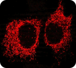
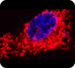
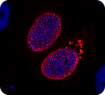
FluoTag®-X4 anti-RFP is a blend of two in-house developed single-domain antibodies (sdAbs) recognizing the most common red fluorescent proteins like mRFP, mCherry, DsRed and other DsRed derivatives with high affinity and specificity. For a complete specificity chart see the Product Specificity Chart below. In our FluoTag-X4 series, two fluorophore molecules are site-specifically coupled to each individual FluoTag molecule. The reagent therefore simultaneously targets up to four fluorophores to the protein of interest, which ensures extra-bright signals. Owing to the small size of our FluoTags, the distance between the target epitope and each fluorophore is below 4 nm. In comparison to conventional detection systems using conventional antibodies, our FluoTag-X4 series can thus improve the localization accuracy by 10-15 nm. Both features – superior brightness and precise fluorophore placement – render our FluoTag-X4 products excellent tools for all microscopy techniques.
- Specificity: Recognizes most common red fluorescent proteins like mRFP, mCherry, dsRed, tdTomato and mScarlet in their native conformation. It does not cross-react with GFP or mTagBFP derivatives. For more detailed information see Product Reactivity Chart below.
- Produced in: E.coli
- Concentration: 2.5 µM protein, 5 µM fluorophore
- Recommended dilution for IF/ ICC: 1:250
- Available fluorophores: Abberior® Star 488, Abberior® Star RED, Abberior® Star 580, Abberior® Star 635P, Atto 488, Atto 542, Atto 580, Atto 647N, Sulfo-Cyanin 3, Sulfo-Cyanin 5. For other fluorophores please contact us.
Relevant Documents
Product List
| PRODUCT NAME | VOLUME | CATALOG NUMBER | ORDER AT |
| FluoTag-X4 anti-RFP | 20 µl | N0404 | Buy online |
| FluoTag-X4 anti-RFP | 200 µl | N0404 | Buy online |
This information is provided without liability and may be subject to change without prior notification.
References
- West et al. (2018) The pro-apoptotic JNK scaffold POSH/SH3RF1 mediates CHMP2BIntron5-associated toxicity in animal models of frontotemporal dementia, Human Molecular Genetics, Volume 27, Issue 8, 15 April 2018, Pages
- Human Molecular Genetics, Volume 27, Issue 8, 15 April 2018, Pages 1382–1395