Description
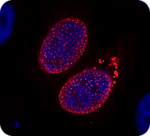
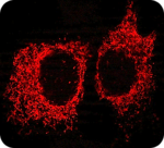
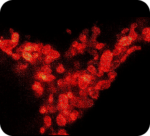
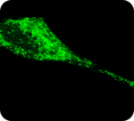
FluoTag®-Q anti-RFP is derived from an in-house developed single-domain antibody (sdAb) recognizing the most common red fluorescent proteins like mRFP, mCherry, DsRed and other DsRed derivatives with high affinity and specificity. For a complete specificity chart see the Product Reactivity Chart below. In our FluoTag-Q series, each fluorophore is coupled to exactly one FluoTag, which in turn binds to its target molecule in a monovalent fashion. The high binding affinity and a high coupling efficiency of > 95% guarantees a highly linear relation between target molecule number and fluorescent intensity. This enables you to directly count your target molecule of interest. The fluorophore is located exceptionally close to the recognized epitope (< 1.5 nm), which is ideal for all microscopy techniques.
- Specificity: Recognizes most common red fluorescent proteins like mRFP, mCherry, dsRed, tdTomato and mScarlet in their native conformation. It does not cross-react with GFP or mTagBFP derivatives. For more detailed information see the Product Reactivity Chart below.
- Concentration: 5 µM protein, 5 µM fluorophore
- Recommended dilution for IF/ ICC: 1:1000
- Available fluorophores: Abberior® Star 488, Abberior® Star RED, Abberior® Star 580, Abberior® Star 635P, Atto 488, Atto 542, Atto 580, Atto 647N, Sulfo-Cyanin 3, Sulfo-Cyanin 5. For other fluorophores please contact us.
Relevant Documents
Product List
| PRODUCT NAME | VOLUME | CATALOG NUMBER | ORDER AT |
| FluoTag-Q anti-RFP | 20 µl | N0401 | Buy online |
| FluoTag-Q anti-RFP | 200 µl | N0401 | Buy online |
This information is provided without liability and may be subject to change without prior notification.